Figure 5.
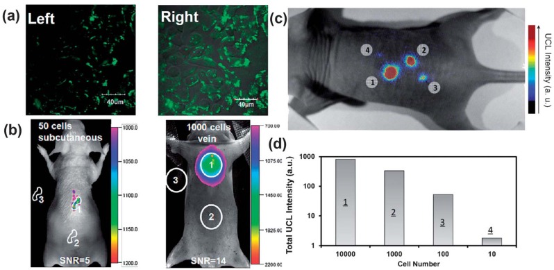
UCNPs for cellular labeling and in vivo tracking analysis. (a) Confocal UCNP imaging (left) and its overlay with a bright field image (right) of cells stained with 200 μg mL−1 NaLuF4 UCNPs for 3 h at 37 °C. (b) In vivo UCNPs imaging of athymic nude mice after subcutaneous injection of 50 human nasopharyngeal epidermal carcinoma KB cells (left) and tail-vein injection of 1000 KB cells (right). The KB cells were pre-incubated with 200μg mL−1 NaLuF4 UCNPs for 3 h at 37 °C before injection. (c and d) In vivo detection of UCNP-labeled mMSCs (an exogenous contast agent to track mouse Mesenchymal Stem Cells). (c) An upconversion luminescence image of a mouse subcutaneously injected with various numbers of mouse mesenchymal stem cells (1 × 10⁵) labeled with UCNPs. (d) Quantification of UCNPs luminescence signals in (c). Adapted with permission from references [87,89], respectively. UCL, upconversion luminescence. Copyright: American Chemical Society, 2011; and Elsevier, 2012.

