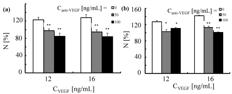
Figure 15.
Proliferation of human umbilical vein endothelial cells triggered by VEGF and neutralization induced by anti-VEGF antibody. HUVECs were first exposed for 2 h to anti-VEGF antibody at two concentrations (Canti-VEGF = 50 and 100 ng/mL). This was followed by the incubation with VEGF (CVEGF = 12 and 16 ng/mL) for (a) 24 and (b) 48 h. The number of cells was normalized to 100% for untreated control cells. *p < 0.01; **p < 0.001(versus cells treated with VEGF only).

