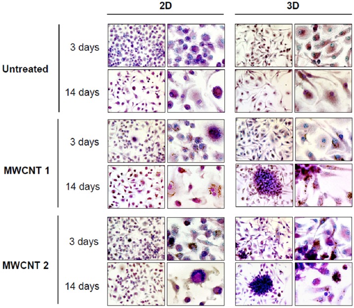
Figure 1.
Light microscopic morphology and kinetics of macrophage aggregation in 2D and 3D cultures. BMDM were exposed to 0.5 μg/mL (0.38 μg/cm2) of particulates. Formation of stable cellular aggregates was evaluated at 3 and 14 days post-exposure. Macrophages were stained with May-Grünwald-Giemsa. Reprinted from [65]. Open Access article, under the terms of Creative Commons Attribution License. Copyright 2011, Licensee Biomed Central Ltd.

