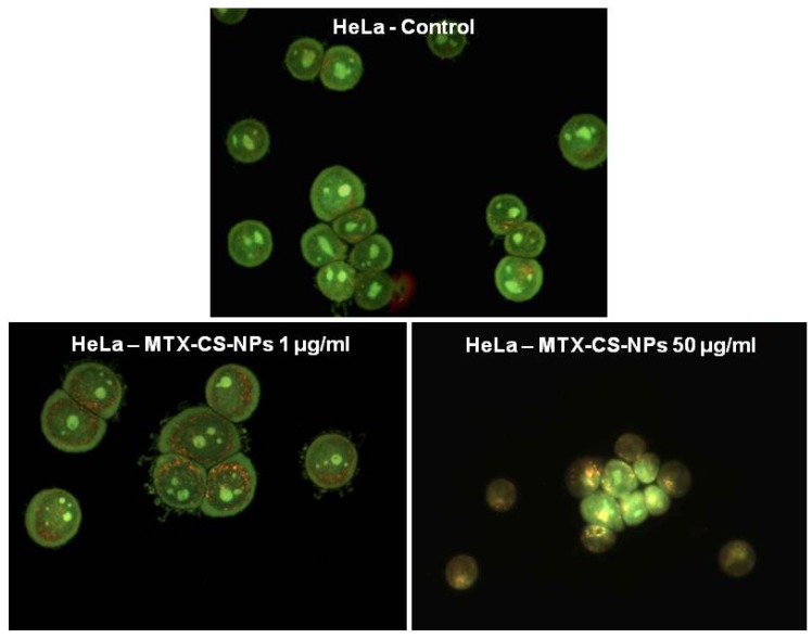
Figure 4.
Assessment of the effects of chitosan NPs encapsulating MTX (MTX-CS-NPs) on lysosomal membrane permeabilization in HeLa cells as visualized via AO staining. In untreated control cells, lysosomes can be seen as red–orange granules and cytoplasm has a diffuse green fluorescence. In cells with lysosomal membrane damage (HeLa cells treated with 50 mg/mL MTX-CS-NPs), lysosomes exhibit a shift from red–orange to a yellow–green fluorescent color. Reprinted with permission from [7]. Copyright 2013, Elsevier.

