Figure 3.
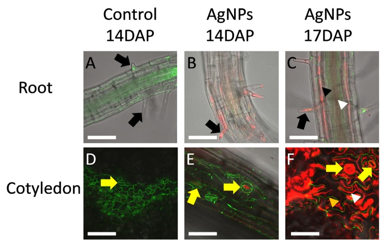
Transport of AgNPs in Arabidopsis ER::GFP plants. Seedlings of 14 (B,E) and 17 (A,C,D,F) days after planting (DAP) were examined under a Zeiss LSM 510 confocal microscope. (A,C) came from root sections of maturation region; (D–F) came from cotyledon. (A) and (D) are control; (B,C) and (E,F) are AgNPs-treated. Green color was intrinsic GFP; red color was Ag0 light scattering. At 14 DAP, AgNPs accumulated mainly in root hair cells and surface of roots (B). By 17 DAP, AgNPs already entered vascular tissue, both phloem (white arrowhead) and xylem (black arrowhead), of the roots and could be bulk transported through vascular tissue. Upon germination, some condensed media might have touched cotyledons. At 14 DAP, AgNPs could be observed in the pores of stomata (yellow arrows in E). By 17 DAP, not only the pores of stomata but also the stomata themselves (yellow arrows in F) showed AgNP accumulation. The uneven surface of pavement cells [56] showed AgNP accumulated on the grooves (white arrowhead) between pavement cells (orange arrow). Scale bar = 0.2 μm.

