Figure 3.
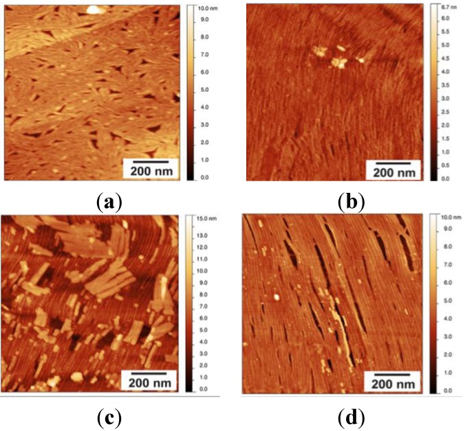
(a) AFM topographic scan of HOPG surface coated with a layer of EAS∆15 protein after a 50-µL drop of EAS∆15 (25 µg/mL) was incubated for 1 min and transferred onto a freshly cleaved HOPG surface; multiple protein rafts are observed; (b) AFM scan of HOPG surface coated with a layer of EAS∆15 protein after a 50 µL drop of EAS∆15 (5 µg/mL) was left to dry overnight onto a freshly cleaved HOPG surface. Sample was imaged directly after drying; (c) a 1-min wash to remove loosely bounded protein layers reveals an underlying, ordered single layer rodlet film; and (d) after 7 min of washing with a stream of running Milli-Q® water (MQW), loose fibril layers are removed and a highly ordered layer of rodlets remains attached to the HOPG.

