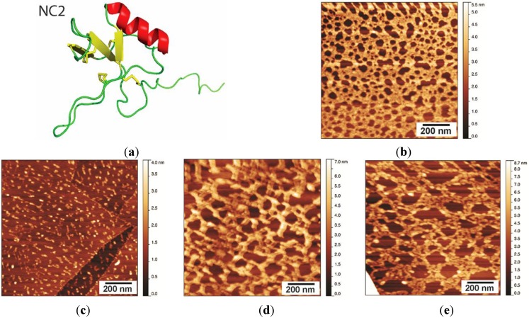
Figure 5.
(a) Ribbon representation of the solution structure of NC2, prepared from PDB Entry 4AOG using PyMol [24]; (b) NC2 layer washed with MQW for 5 min, displaying a protein network with pores of 20–30 nm and a layer height of 1.5–2 nm; (c) NC2 layer after treatment with 60% ethanol for 5 min; (d) NC2 layer after treatment with 3 M NaOH; and (e) NC2 layer after treatment with 3 M HCl.

