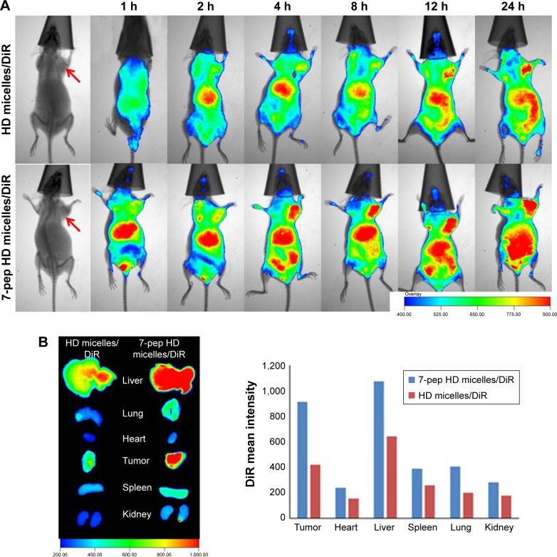
Figure 7.
In vivo and ex vivo imaging tests.
Notes: (A) The fluorescent and X-ray pictures of nude mice bearing MCF-7/Adr tumors at various time points after administration with HD micelles/DiR and 7-pep HD micelles/DiR via tail vein. The red arrow points out the tumors in each mouse. (B) Ex vivo fluorescent images and qualitative data of tumors and main organs excised from the mice at 24 h after injection.
Abbreviations: DiR, 1,1-Dioctadecyl-3,3,3,3-tetramethylindotricarbocyanine iodide; h, hour; HD micelles, PHIS-PEG2000 and DSPE-PEG2000 hybrid micelles; DSPE-PEG2000, 1,2-distearoyl-sn-glycero-3-phosphoethanolamine-polyethylene glycol-2000; PHIS-PEG2000, poly(l-histidine)-coupled polyethylene glycol-2000; 7-pep HD micelles, PHIS-PEG2000 and 7pep-DSPE-PEG2000 hybrid micelles; 7pep, HAIYPRH, a TfR ligand.

