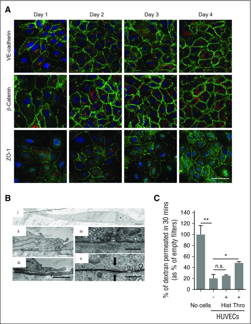
Figure 2.
Confluent HUVECs on polyester membranes form a polar monolayer. (A) Immunofluorescence of confluent HUVECs grown on polyester membranes for the times indicated (days) and stained for the cell’s nucleus (DAPI, blue); VWF (red); and VE-cadherin, β-catenin, or ZO-1 (green). Scale bar represents 50 μm. (B) TEM of a cross-section of a HUVEC (i) with the TGN to the right of the nucleus (*). Two examples of TEM cross-sections of overlapping tips between neighboring HUVECs (ii-iii) and the close contact formed between the cell membranes of 2 adjacent cells (iv-v) with “fuzz”-like structure typical of tight junctions in epithelial cells (arrows). Scale bar: i, 2 μm; ii-iii, 1 μm; iv-v, 300 nm. (C) Dextran permeability assay, where 1 mg/mL of 40 kD-FITC-dextran was loaded on the apical chamber of empty membranes (No cells), 2-day confluent unstimulated HUVECs (-), or HUVECs treated with 100 μM histamine (+Hist) or 2.5 U/mL thrombin (+Thro) to open extracellular pores. The amount of FITC-dextran in the basolateral chamber was measured after incubation with FITC-dextran in the apical chamber for 30 minutes. Mean of triplicate wells (with standard deviation [SD]), as percent of No cells control sample. *P < .05; **P < .005; n.s., not significant.

