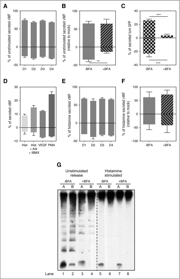
Figure 3.
Polarity of VWF secretion. (A) Unstimulated VWF secretion on Transwell membranes of HUVECs grown for the time indicated (in days), expressed as a percent of secreted VWF from the total VWF in each sample (secreted plus lysate). Results standardized to D1 sample. Representative experiment, mean of 3 replicates with SD. (B) Unstimulated VWF secretion from HUVECs grown on Transwell membranes for 2 days. Cells were either left untreated (-BFA) or were treated with 5 μM of Brefeldin A (+BFA) for 1 hour before and during secretion collection from both chambers. Results standardized to control -BFA sample. Mean of 3 independent experiments with standard error of the mean (SEM). (**P < .005). (C) lumGFP secretion on Transwell membranes. HUVECs were nucleofected with lumGFP construct and seeded on Transwell membranes for 30 hours before secretion assay. Cells were treated with either Control (-BFA) or 5 μM of Brefeldin A (+BFA) for 1 hour before and during secretion collection from both chambers. Amount of lumGFP secreted expressed as a percent of total GFP in each sample (secreted plus lysate). Representative experiment reflects the mean of 3 replicates with SD. (***P < .0005). (D) Stimulated VWF secretion from HUVECs grown on Transwell membranes for 2 days. Cells were stimulated with either 100 μM histamine (Hist), 100 μM histamine plus 100 μM adrenaline plus 100 mM IBMX (Hist +Adr +IBMX), 40 ng/mL vascular endothelial growth factor, or 100 ng/mL PMA for 30 minutes while secreted VWF was collected from both chambers. Amount of VWF secreted expressed as a percent of total VWF in each sample (secreted plus lysate). Representative experiment reflects the mean of 3 replicates with SD. (E) Histamine VWF secretion from HUVECs grown on Transwell membranes for the time indicated (in days), expressed as a percent of total VWF in each sample (secreted plus lysate), relative to control (D1) sample. Results standardized to D1 sample. Representative experiment, mean of 3 replicates with SD. (F) Histamine VWF secretion from HUVECs grown on Transwell membranes for 2 days. Cells were treated with either control (-BFA) or 5 μM of Brefeldin A (+BFA) for 1 hour before and during 30 minutes 100 μM histamine stimulation and secretion collection from both chambers. Results standardized to control -BFA samples. Mean of 3 independent experiments with SEM. (G) Representative multimer pattern of secreted VWF collected from unstimulated and histamine-stimulated HUVECs. Cells were treated with either control (-BFA) or 5 μM of Brefeldin A (+BFA) for 1 hour before and during VWF secretion collection in both chambers. Unstimulated secretion was collected for 1 hour and 100 μM histamine stimulated secretion was collected for 30 minutes. The same units of VWF (as measured by ELISA) were loaded for all samples. VWF ELISA quantification corresponding to these samples is shown in (B) and (F). A, apical chamber; B, basolateral chamber. Lanes 1-4, unstimulated releasate; lanes 5-8, histamine-stimulated releasate.

