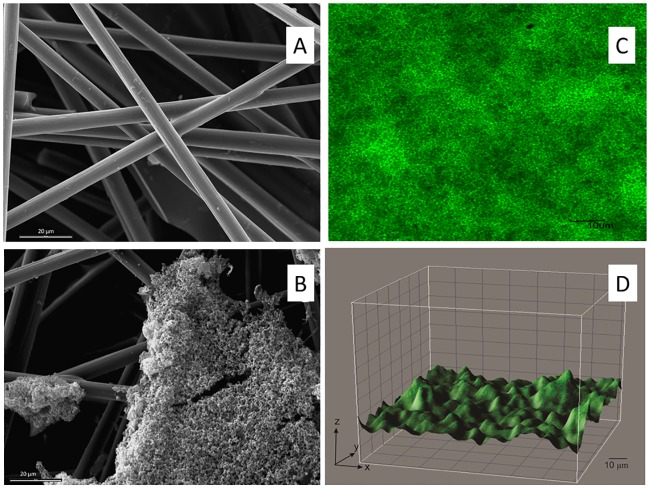
Fig 5. Microphotography of the working electrode used in the BES in order to understand the morphology of the biofilm formed by Dietzia sp. RNV-4 on carbon paper electrodes.
(A) Electrode from a control experiment, 1000X, bar 20 μm, by SEM. (B) Biofilm of Dietzia sp. RNV-4, 1000X, bar 20 μm, by SEM. (C) Biofilm of Dietzia sp. RNV-4, 60X zoom 2.5, bar10 μm, by CLSM. (D) Biofilm of Dietzia sp. RNV-4, 60X zoom 2.5, by confocal microscopy. XYZ images were processed using the program Image J.

