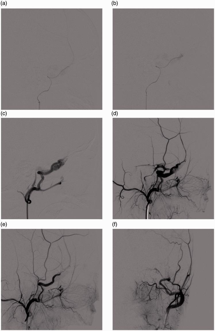
Figure 3.
DSA images following treatment. (a) The microcatheter crossed the fistula orifice, and the angiography clearly shows the MMA; (b) The microcatheter was introduced into the fistula orifice, and the angiography shows the PP. (c) Lateral external carotid artery angiography indicates embolization with coils in the fistula orifice. (d) After the coil embolization, Onyx gel was injected to fill the AVF. (e) and (f) AP and lateral angiography at the end of the embolization indicates the disappearance of the AVF and the re-development of the distal end of the MMA. DSA: digital subtraction angiography; MMA: middle meningeal artery; PP: pterygoid plexus; AVF: arteriovenous fistula; AP: anteroposterior.

