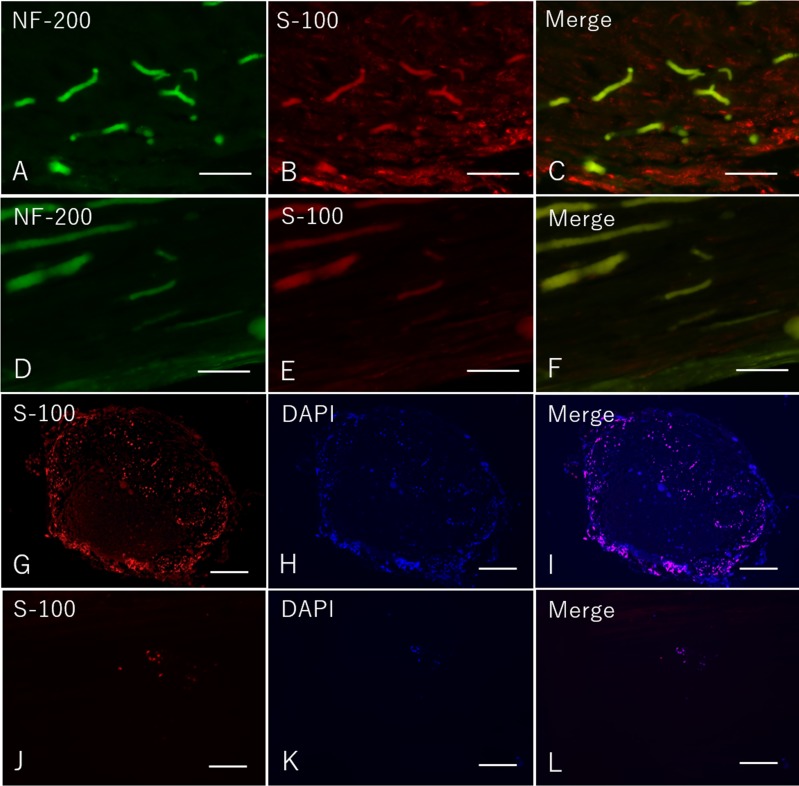
Fig 6. Immunohistochemistry of the mid portion of the regenerated nerve eight weeks after surgery in both groups.
A-C: Longitudinal sections in the Bio 3D group. D-F: Longitudinal sections in the silicone group. G-I: Transverse sections in the Bio 3D group. J-L: Transverse sections in the silicone group. A-F: scale bar = 100 μm. G-L: scale bar = 500 μm.

