Figure 1.

Detection of the neuromas.
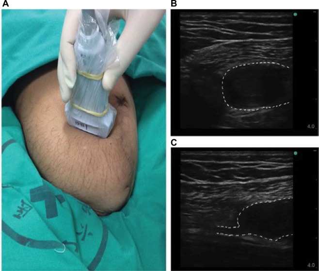
Notes: (A) Using ultrasound probe to scan the stump limb. (B) The transverse axial view of neuroma. (C) The longitudinal axial view of neuroma. The dotted line indicates neuroma.

Detection of the neuromas.
Notes: (A) Using ultrasound probe to scan the stump limb. (B) The transverse axial view of neuroma. (C) The longitudinal axial view of neuroma. The dotted line indicates neuroma.