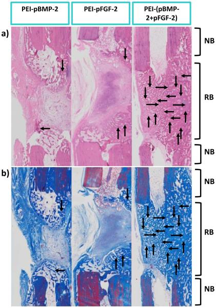
Fig.6.
Images of histology sections indicating the degree of nascent bone formation at the defect sites at 28 days in response to indicated treatments. Representative histology sections after (a) H&E, and (b) Masson’s trichrome staining. As shown, there is qualitatively more bone area in the combination PEI-(pBMP-2+pFGF-2) group compared to the PEI-pBMP-2 or PEI-pFGF-2 only. NB-native bone and RB-regenerated bone. Note the complete bridging of new bone (indicated by the arrows) in the group treated with scaffolds embedded with PEI-(pBMP-2+pFGF-2) nanoplexes.

