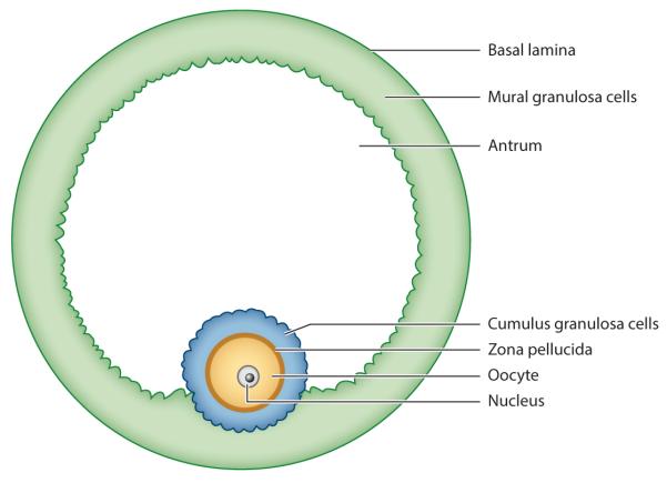
Figure 2.
Tissue layers of a mammalian preovulatory follicle. The oocyte with its prophase-arrested nucleus is surrounded by 2–3 layers of cumulus granulosa cells, which are attached in one region to the 5–10 layers of mural granulosa cells. Elsewhere, a fluid-filled antrum separates the two types of granulosa cells.

