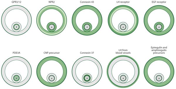
Figure 3.
Localization of some of the signaling proteins that regulate meiotic arrest and resumption in preovulatory follicles. Protein distribution is either determined directly, by ligand binding, immunofluorescence, or Western blotting; or it is inferred from mRNA distribution, by in situ hybridization, or from RT-qPCR. Green indicates the presence of the protein, and lighter green indicates a lesser amount of the protein. White indicates that the protein (or mRNA) was either not detected or detected at a level ≤10% of that elsewhere. Figure is based on data from the following references: CNP precursor (66); connexin 43 (9), connexin 37 (9), EGF receptor (99; LA Jaffe and JR Egbert, unpublished data); epiregulin and amphiregulin precursors (99); GPR3/12 (40, 46); LH from blood vessels (14); LH receptor (84); NPR2 (66, 70); and PDE3A (61, which used a previous nomenclature, PDE3B). Abbreviations: CNP, C-type natriuretic peptide; EGF, epidermal growth factor; LH, luteinizing hormone; NPR2, natriuretic peptide receptor 2; PDE3A, phosphodiesterase 3A.

