Fig. 1.

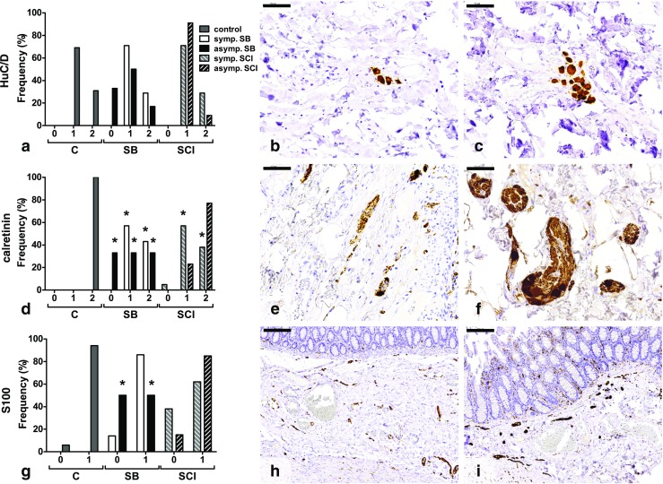
Histological findings in the submucosal plexus of colon. a No significant differences were found between the HuC/D-stained sections. Most tissues showed low neuronal densities (grade 1) (b), while in some sections, high neuronal densities (grade 2) were found (c). d Significantly less calretinin-positive neurons were found in both SB groups and the symptomatic SCI group compared with the control group. e Section with neurons present, but less than one neuron per neuronal structure (grade 1). f Minimal one neuron per neuronal structure is shown in this tissue (grade 2). g Low nerve fiber densities (grade 0) were most often observed in the asymptomatic SB group. h, i Illustration of low (grade 0) (h) and high (grade 1) (i) nerve fiber density. Numbers on the x-axis represent the semiquantitative scores. C control, SB spina bifida, SCI spinal cord injury. *p < 0.05 vs control. Scale bars 50 μm (b, c, e, f) and 200 μm (h, i)
