
Fig. 1.
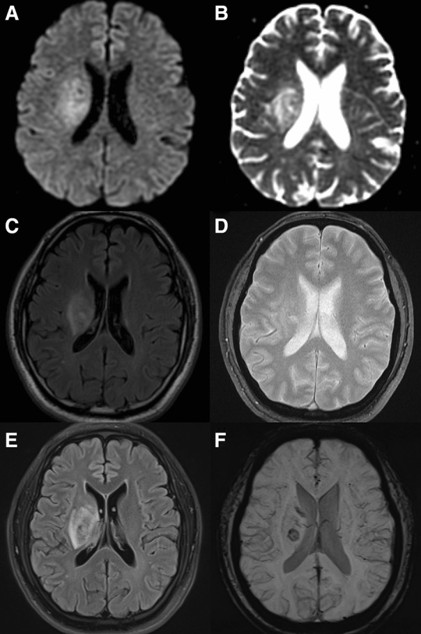
Initial and follow-up magnetic resonance images (MRI) at the first event. The initial MRI shows high signal intensity on diffusion-weighted image (DWI, a), without low signal intensity on apparent diffusion coefficient (ADC, b). Fluid-attenuated inversion recovery (FLAIR, c) images reveal increased signal intensities in right basal ganglia and centrum semiovale. No hemorrhagic lesion was detected in gradient-echo images (GRE, d). The follow-up MRI shows slightly increased extent of FLAIR high signal intensity area (e) and development of focal hemorrhage at right basal ganglia (f), compared to the initial MRI images