Fig. 5.
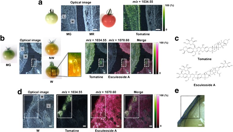
Visualization in more local physiological responses to ripening and wounding stress with DHB-based MALDI–MSI. a Optical image and ion image of tomatine in thin-sections of between MG and MR tomato fruits. M mesocarp, L locule. b Optical image and ion images derived from tomatine-related compounds among three phenotypes of tomato fruits, MG, non-wounded (NW), and wounded (W). Both NW and W fruits in pink stage, corresponding to the mid-stage of ripening. c Structures of tomatine (m/z = 1034.55 [M + H]+) and esculeoside A (m/z = 1270.60 [M + H]+), a glycosylated tomatine product. d Optical image and magnified ion images derived from tomatine-related compounds focused on the wound region. e Evans Blue staining for viability of the cells around wound region in tomato fruit. Blue-colored area; non-viable cells. The broken box; wound region. Scale bar = 1.0 mm

