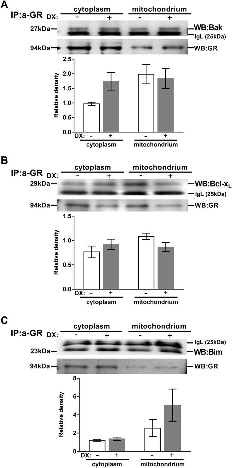
Fig. 2.
Association of the GR with members of the Bcl-2 family in thymocytes. Anti-Bak (a), Bcl-xL (b) and Bim (c) western blots are shown from cytoplasmic and mitochondrial fractions of thymocyte lysates after anti-GR precipitation with or without DX treatement. Blots were reprobed with anti-GR antibody to confirm equal loading of the samples. The figure shows representative blots and densitometry data of at least three independent experiments. Diagrams below each blot show the relative Bak, Bcl-xL and Bim levels in the cytoplasm (normalized to GR) and the mitochondria (normalized to GR). Bars represent the mean ± SEM of relative densities compared with the controls. IgL: immunoglobulin light chain

