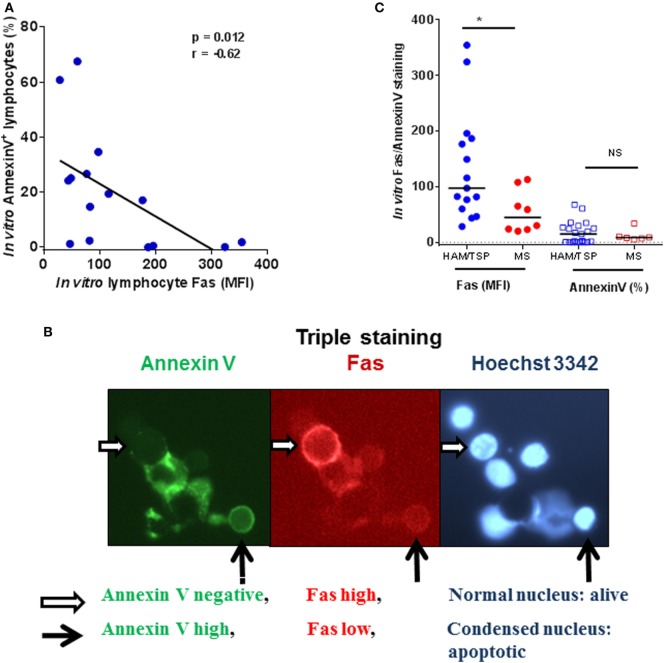
Figure 5.
Fashi cells are apoptosis-resistant in HAM/TSP patients. (A) Fas mean fluorescence intensity (MFI, on a per-cell basis) negatively correlates to apoptosis (quantified as % annexin V+ cells) in lymphocytes of HAM/TSP patients (*p = 0.012, Spearman’s r = −0.63, n = 15). (B) In the middle panel is a representative image of a non-apoptotic Fashi cell (indicated by a white horizontal arrow). This Fashi cell is annexin V negative as visualized in the first panel and displays a normal nuclear morphology seen in the third panel. On the contrary, a Faslo cell in panel 2 (black vertical arrow), displays pronounced annexin V staining (panel 1) and is undergoing apoptosis, as evidenced by nuclear condensation, and is being engulfed by a macrophage (panel 3). (C) In vitro Fas levels (MFI) and apoptosis (% of Annexin V+ cells) are compared between neuroinflammatory diseases HAM/TSP and multiple sclerosis (Mann–Whitney test, *p < 0.05).

