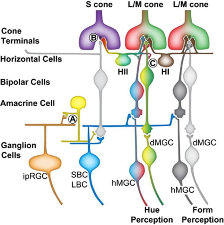
Figure 2.
Three different types of synaptic mechanisms provide S-cone input to a diversity of primate ganglion cells. (a) Glycinergic inhibitory S-cone amacrine cells are proposed to be responsible for both transmitting and sign inverting S-ON-bipolar cell signals to melanopsin ganglion cells allowing changes in the color of light to contribute cues to circadian activity. (b) Synapses between S-cones and S-cone specific ON-bipolar cells are responsible for small and large bistratified ganglion cells having cone inputs configured as S-(L+M). (c) In the retina, L/M opponent cells with S-cone inputs could result from GABA-mediated feedforward from S-cones to L/M midget bipolar cells. L/M opponent cells with S-cone inputs input have been observed in four different configurations: (S+M)-L, L-(S+M), (S+L)-M, and M−(S+L), exactly the same as the mechanisms known to underlie conscious hue perception. Midget ganglion cells without S-cone input carry color information but they also are extremely well suited to mediating very-high acuity achromatic spatial vision. In ancestors to modern primates in which the majority of individuals were dichromatic, the homologs to the L/M opponent cells can only serve achromatic vision. Similarly, L/M opponent cells without S-cone inputs may only function to serve achromatic form vision in trichromats. Abbreviations: dMGC, depolarizing midget ganglion cell; hMGC, hyperpolarizing midget ganglion cell; ipRGC, intrinsically photosensitive ganglion cell; LBC, large bistratified ganglion cell; SBC, small bistratified ganglion cell.

