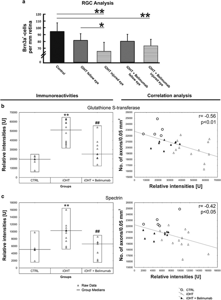
Figure 2.
Analysis in intermittent ocular hypertension model (iOHT). (a) The survival of RGC was analyzed as the number of Brn3a-positive cells per mm retina in naso-temporal cross-sections of the eye in control animals (CTRL), animals that received unilateral intermittent ocular hypertension (iOHT), and animals with unilateral intermittent ocular hypertension which received B lymphocyte inhibitor Belimumab treatment (iOHT+Belimumab). A significant loss of RGC was observed in injured eyes of iOHT group and iOHT+B lymphocyte inhibitor Belimumab group, to hinder B lymphocyte activation, (both P<0.01, horizontal lines) compared with control eyes (black). Furthermore, a significant difference in damage was observed between fellow and injured eye of iOHT group (P<0.05. Significant values are indicated as follows: *P<0.05, **P<0.01). (b and c) Quantification of different antigen reactivities The left side shows the immunoreactivities in relative intensities per group against glutathione S-transferase (b) and spectrin (c). Each triangle represents one animal, and the black line indicates the group median. Compared with the relative intensity of CTRL, iOHT was significantly upregulated (**P<0.01) for all investigated antigens. Compared with iOHT, all immunoreactivities of iOHT+Belimumab were significantly downregulated (##P<0.01). Scatterplots of the number of optic nerve axons in a distinct area (no. of axons/0.05 mm2) against relative intensities of glutathione S-transferase (b) and spectrin (c), are shown on the right side. The gray fitting line shows the negative linear dependence between the number of axons per 0.05 mm2 and the relative intensities. Significant values are indicated as follows: **P<0.01 compared with control group, ##P<0.01 compared with iOHT group. (Adapted from Gramlich et al43)

