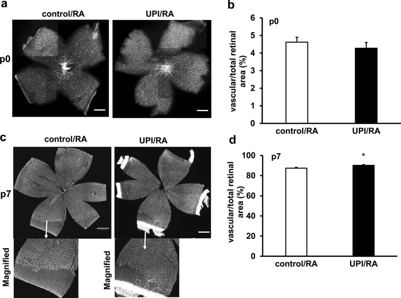
Figure 2. UPI pups show increased physiological retinal vascularization in room air (RA).
(a) Representative retinal flatmount images (Scale bar, 990 μm) and (b) quantification of percent of vascularized retinal area/total retinal area at postnatal day 0 (p0); (c) Representative retinal flatmount images (Upper row-Mag, 4X and lower row-Magnified images; Scale bar, 990 μm) and (d) quantification of percent of vascularized retinal area/total retinal area at p7 (*p < 0.05 vs. control/RA; n = 9–32).

