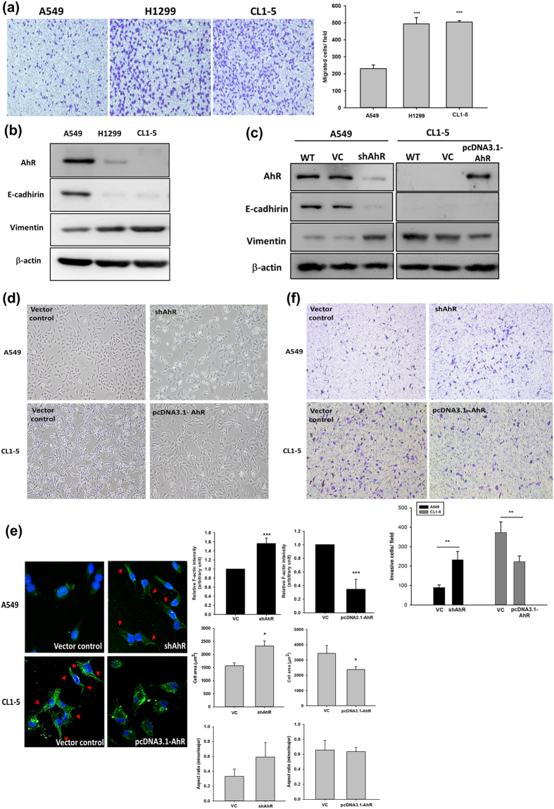
Figure 1. Aryl hydrocarbon receptor (AhR) expression in different cells was correlated with E-cadherin and vimentin expression and cell motility.
Protein expression was evaluated by western blotting. Cells (104 cells/Transwell) were seeded on matrigel-coated Transwell inserts and incubated for 16 h. The migrated/invasive cells were stained with crystal violet and counted using the Image-Pro plus software. Images were acquired at 40× magnification. (a) Cell migration potential between A549, H1299, and CL1-5 cells. (b) Expression of AhR, E-cadherin, and vimentin in A549, H1299, and CL1-5 cells. Full-length blots are presented in Supplementary Fig. S11. (c) AhR silencing and overexpression in A549 and CL1-5 cells, respectively, and vimentin expression but not E-cadherin was also affected. Full-length blots are presented in Supplementary Fig. S12. (d,e) Cell morphology was altered in A549 and CL1-5 cells. Immunostaining of F-actin (green fluoresce) was observed by confocal microscope. The fluorescence intensity (a.u.), cell area and minor/major axis ratios were also measured by Image ProTM and data analysed as indicated. Arrowheads indicate cells expressing the actin stress fibers. (f) And the cell invasive potential were altered in A549 and CL1-5 cells. The quantified data were analysed as cell number per field and expressed as the mean ± SD from three independent experiments. **P < 0.01; ***P < 0.001 compared to the control group.

