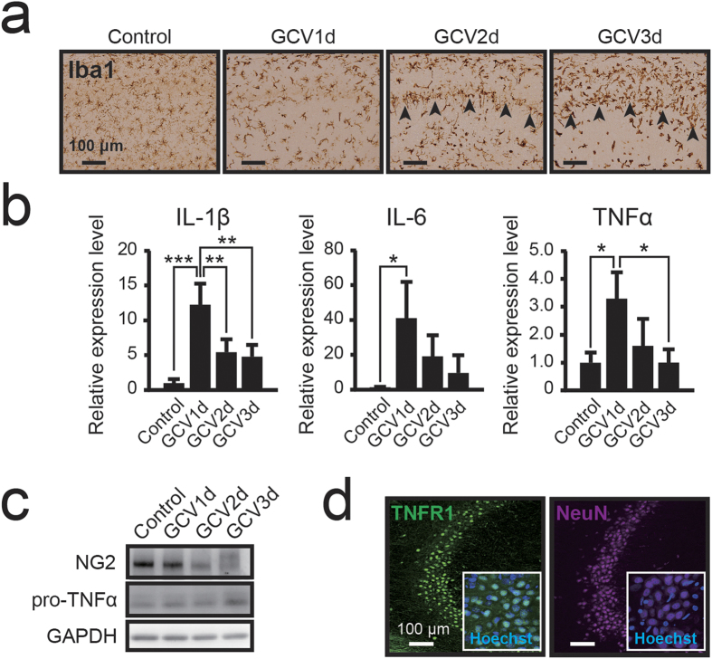
Figure 4. Ablation of NG2 glial cells induces neuroinflammation and activates microglia in the hippocampus.
(a) Bright-field immunohistochemical observations of Iba1 in NG2-HSVtk transgenic rats treated with vehicle (Control) or GCV for 1, 2, or 3 days (GCV1d, GCV2d, GCV3d). Black arrowheads indicate the CA1 pyramidal cell layer. (b) Relative expression levels of interleukin-1β (IL-1β), interleukin-6 (IL-6), and tumor necrosis factor α (TNFα) mRNAs in the hippocampal tissue of NG2-HSVtk transgenic rats treated with vehicle or GCV for 1, 2, or 3 days. (c) Immunoblotting of NG2 and precursor protein of TNFα (pro-TNFα) in the hippocampal tissue of NG2-HSVtk transgenic rats treated with vehicle or GCV for 1, 2, or 3 days. Glyceraldehyde 3-phosphate dehydrogenase (GAPDH) was used as a loading control. (d) Confocal images (magnified views in white boxes) of immunoreactivity for TNF receptor 1 (TNFR1) and NeuN in the hippocampal CA2 region in NG2-HSVtk transgenic rats. Mean ± SD, n = 3 rats (Control, GCV3d) and 4 rats (GCV1d, GCV2d); *p < 0.05, **p < 0.01 or ***p < 0.001, based on a one-way ANOVA followed by Tukey-Kramer test. Scale bars represent 100 μm (a,d).

