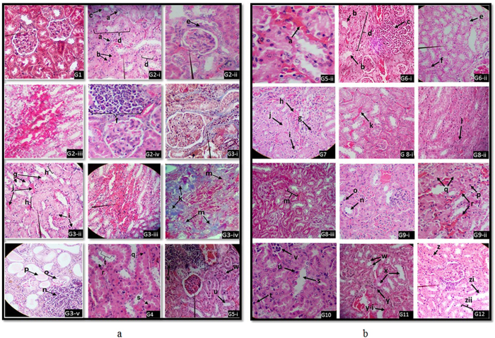
Figure 1.
Histopathological evaluation of renal tissues in; (a) G1-Intact tubules, pointer-glomeruli and interstitium (H&E × 100). G2-i Tubular interstitial lesions, a-Formation of fibrous bands, b-Apoptotic cells with nuclear pyknosis, c- Tubular atrophy in proximal convoluted tubule (PCT), d, Pointer – Degeneration and necrosis of PCT (H&E × 100). G2-ii-Initiation of glomerular sclerosis, e-Fibrosis of Bow man capsules. Pointer–thicken Bow man capsule (H&E × 100). G2-iii-Hemorrhages at cortico-medullary area (H&E × 100). G2-iv-Interstitial inflammatory cell infiltration at renal cortex, f-infiltrated mononuclear inflammatory cells (H&E × 100). G3-i -interstitial fibrosis (blue colour, pointer) (MT × 100). G3-iiTubular lesions, g-Tubular atrophy (loss of brush borders with clear lumen in PTC, h-Degenerated and necrotic areas of PCT, i-Apoptosis of tubular epithelial cells with pyknotic nuclei, j-Hyperemic renal capillaries (H&E × 100). G3-iii-pointer-hemorrhagic area (H&E × 40). G3-iv-k-Global glomerular sclerosis (Blue colour), m-interstitial fibrosis (blue colour) (MT × 100). G3-v-Reduced functional renal tissue density and interstitial fibrosis, n-Mononuclear inflammatory cell infiltration, o-Tubular atrophy in PCT, p-Protein cast in a tubular lumen (H&E × 100). G4-Tubular lesions, q-Apoptotic cells with pyknotic nuclei PCT, r-Tubular atrophy (brush borders are lost with clear lumen) in PCT, s-degenerated tubular epithelial cell in a PCT, t-Necrotic area of a PCT (H&E × 100). G5-i,u-Tubular atrophy. v-Cell infiltration, w–Apoptotic cell with nuclear pyknosis, pointer-intact glomerulus (H&E × 40). (b) G5-ii, a-Hyperemic capillaries (H&E × 100). G6-i, b-Tubular atrophy in PCT (loss of brush borders with clear lumen), c-Mononuclear inflammatory cell infiltration, d-replacement of renal tissue with fibrous tissue in renal cortex (H&E × 100). G6-ii, pointer–PCT undergoing atrophy (loss of brush borders with clearing of the lumen) e-Degeneration and necrosis of tubules, f-Apoptotic cell with nuclear pyknosis (H&E × 40).G7, g-Apoptotic tubular epithelial cell, h-Hyperemic capillaries at cortical area, i-Initial stages of fibrosis, j-Degeneration and necrosis of PCT (H&E × 40). G8-i, k-Hyperemic capillaries (H&E × 100).G8-ii,l-Apoptotic cells of a PCT. G8-iii, m-Tubular degeneration and necrosis (MT × 40). G9-i, n-Tubular atrophy of a PCT, o-Apoptotic cells with nuclear pyknosis, (H&E × 100). G9-ii, p- Apoptotic cell. Q-Degeneration and necrosis of PCT, r-Hyperemic capillaries. (H&E × 100). G10, s-Tubular atrophy, t-Apoptosis, u-Degenerated cell of a PCT, v-Mononuclear cell infiltration (H&E × 100). G11, w-Apoptotic tubular epithelial cell, x-Tubular atrophy of PTC, y-Interstitial fibrosis, y-i-Hyperemic capillaries (H&E × 40).G12, z- Apoptotic cell, zi-Tubular atrophy of a PCT, Pointer-Mononuclear cell infiltration, zii-Cell degeneration and necrosis (H&E × 100).

