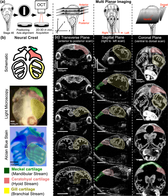
Figure 3.
Imaging Xenopus craniofacial structures with OCT (a) We create a slit at the center of the clay with the Stage 46 tadpole positioned ventrally towards the OCT imaging plane (XZ-plane). Then we adjust the 5-axis stage to capture a zero- degree reference axis where a plane intersects the tadpole’s eyes symmetrically and the tip of the tail. This plane defines the 0° orthogonal plane. We then move this imaging plane in y, x and z axes to capture transverse, sagittal and coronal sections respectively. Imaging planes are adjusted on all three axes to capture distinct facial structures. (b) Top panel shows the schematic representation of the three neural crest streams: mandibular (green), hyoid (pink) and branchial (yellow) which form Meckel, ceratohyal and gill cartilages respectively. Each structure is highlighted under simple stereomicroscopy image (middle panel) and after alcian blue stain (bottom panel) (c) OCT images of craniofacial structures in all three planes: Transverse, Sagittal and Coronal. Meckel’s cartilage (green label) is best viewed in sagittal (yz-axis) and coronal (xy-axis) sections. The ceratohyal cartilage can be easily resolved by the coronal sections. The gill cartilages are more posterior compared to Meckel’s and ceratohyal cartilage, and the gill cartilages occupies most of the craniofacial space. Qualitative analyses can be made in all axes.

