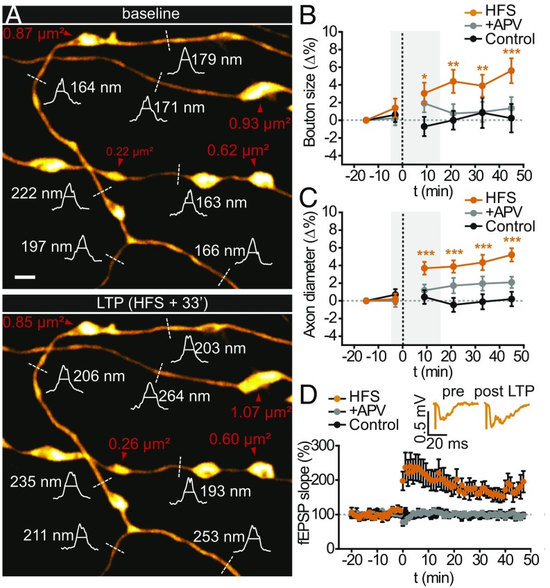
Fig. 3.
STED imaging of long-term dynamics of synaptic boutons and axon diameters. (A) Synaptic boutons (red arrowheads) remained slightly enlarged, and axon diameters (white curve, intensity profiles; dashed lines, FWHM) grew wider relative to pre-HFS baseline levels. (B and C) Quantification of late morphological changes. (B) Normalized synaptic bouton sizes for HFS, control, and APV conditions. (C) Normalized axon diameters for these conditions. The gray boxes in B and C correspond to the time window of the short-term experiments in Fig. 2. (D) Normalized time course of the fEPSP slope during LTP induction. Inset shows representative fEPSP traces. LTP was blocked by APV, and the recordings were stable under control conditions. (Scale bar, 1 µm.)

