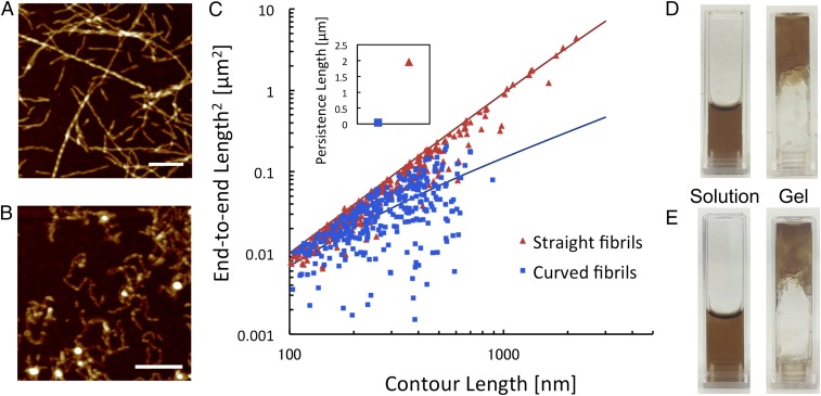
Fig. 1.
Two distinct morphological classes of PNFs formed from WPI. (A and B) AFM images of straight (A) and curved (B) fibrils prepared from initial WPI concentrations of 4% and 8%, respectively. [Scale bars: 1 μm (A) and 0.3 μm (B).] (C) Plot of the squared end-to-end distance versus contour length for straight and curved PNFs with the best-fit curves used to calculate the persistence lengths. (Inset) Final values of persistence lengths for straight and curved fibrils. (D and E) Photographs of PNF dispersions, before (Left) and after (Right) the addition of acetate buffer (pH 5.2) for straight fibrils (D) and curved fibrils (E).

