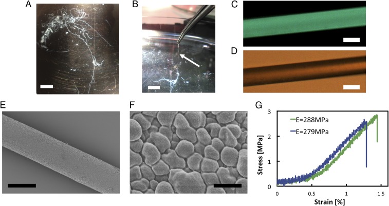
Fig. 3.
Characterization of microfibers obtained from curved PNFs. (A) Photograph of a hydrogel fiber in the bath solution after spinning in the flow-focusing setup. (B) The fibers made from curved PNFs can be picked up from the bath. (C) Fluorescence microscope image of a fiber stained with thioflavin T. (D) Optical microscope image of the fiber shown in C. (E) SEM image of a dried fiber. (F) Zoomed SEM view of the fiber surface. (G) Representative stress–strain curves of dried fibers. [Scale bars: 1 cm (A and B), 30 μm (C–E), and 1 μm (F).]

