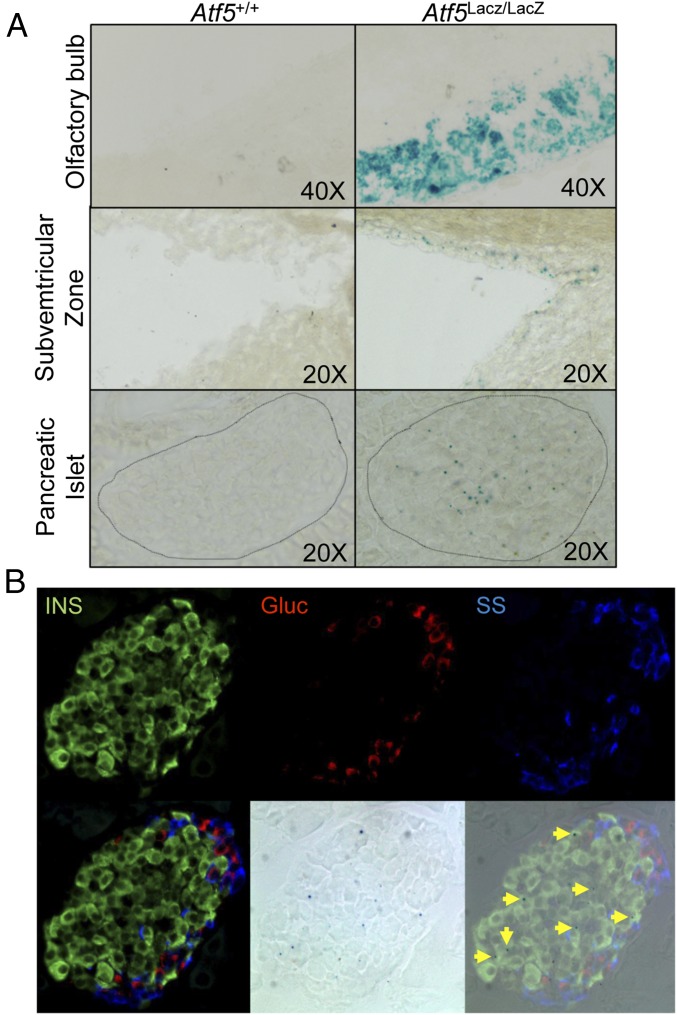
Fig. 1.
ATF5 is expressed in pancreatic islets. (A) β-Gal activity assessed by X-Gal staining of the olfactory bulb (Top), subventricular zone (Center), and islets (Bottom) from Atf5+/+ (Left) and Atf5LacZ/LacZ (Right) mice. (B) β-Gal and immunofluorescent staining of parallel sections from Atf5LacZ/LacZ pancreata for insulin (Upper Left), glucagon (Upper Center), and somatostatin (Upper Right). The lower row shows merged (Lower Left), β-gal bright-field (Lower Center), and merged IF and X-Gal (Lower Right) images. Arrows indicate insulin-positive, β-gal+ cells.

