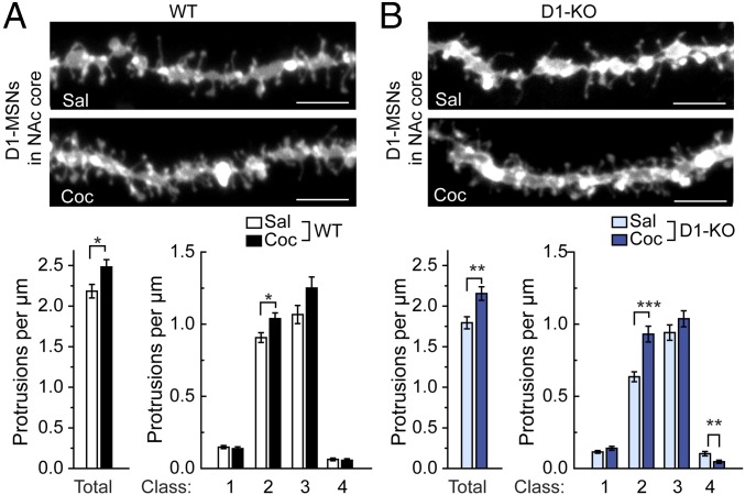
Fig. 4.
Chronic cocaine-induced changes in dendritic spine density. WT mice (A) and WAVE1 D1-KO mice (B) were treated daily (i.p.) with saline (Sal) or cocaine (Coc) (15 mg/kg) for 14 d. Two days after the last injection, mice were perfused and dendritic spines were analyzed in DiI-labeled D1-MSNs (identified as enkephalin− cells) in the NAc core. (Upper panels) Representative images of dendritic segments. (Scale bars, 5 µm.) (Lower graphs) Quantification of total and four classes of dendritic protrusion density. Class 1 represents stubby spines without a discernible neck, classes 2 and 3 represent short and long spines, respectively, and class 4 represents filopodial protrusions. Data represent means ± SEM. *P < 0.05, **P < 0.01, and ***P < 0.001, Kolmogorov–Smirnov test. n = 14–16 mice per group. The total numbers of dendrites analyzed were 36 (WT-Sal), 30 (WT-Coc), 45 (KO-Sal), and 30 (KO-Coc).

