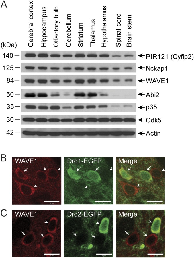
Fig. S1.
Expression of WAVE1 complex and Cdk5/p35 in the brain and expression of WAVE1 in D1- and D2-MSNs. (A) Immunoblotting of the components of the WAVE1 complex, Cdk5, p35, and actin in brain regions as indicated. (B and C) Immunohistochemically stained WAVE1 and EGFP in the striatum of Drd1a-EGFP mice (B) or Drd2-EGFP mice (C). Arrows and arrowheads indicate D1-MSNs and D2-MSNs, respectively. WAVE1 expression is observed in both D1-MSNs and D2-MSNs. (Scale bars, 10 µm.)

