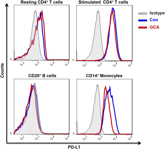
Fig. S2.
PD-L1 expression on T cells, B cells, and monocytes in GCA patients. PBMC were collected from GCA patients with active vasculitis. Naïve CD4+ T cells were isolated and stimulated with anti-CD3/CD28 beads for 7 d. Cells were stained with anti-CD4, anti-CD20, anti-CD14, and anti–PD-L1 Ab. Data were acquired by flow cytometry. Representative flow charts are shown.

