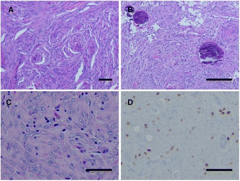
Fig. 2.

Histological appearance of benign and atypical meningioma. a Histological analysis of the posterior specimen revealed a grade I meningioma with typical whorl formations (bar 100 μm) and b psammoma bodies (bar 100 μm). c The anterior tumor revealed a grade II meningioma with increased cellularity, nuclear pleomorphism, and prominent nucleoli as indicated by H&E (bar 50 μm) and d an increased proliferation index by MIB-1 labeling (bar 50 μm)
