Fig. 2.

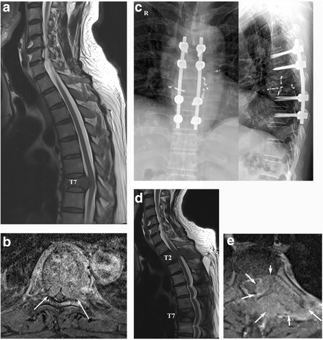
A 54-year old man with HCC who developed MSCC. a Preoperative T2-weighted sagittal MR image showing cord compression at T7. b Preoperative T1-weighted enhanced MR image (arrows indicate cord compression by the tumor mass). c Postoperative radiographs. d T2-weighted sagittal MR image 6 months postoperatively showing another occurrence of cord compression at T2, with maintenance of the decompression at T7. e T1-weighted enhanced MR image at T2 (arrows indicate tumor mass). Hepatocellular carcinoma (HCC), metastatic spinal cord compression (MSCC)
