
Fig. 1.
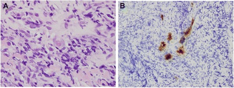
a Cytomegalovirus in an endothelial cell (hematoxylin and eosin stain, 1000× magnification). b Immunohistochemistry of cytomegalovirus-infected cells (400× magnification)
Official websites use .gov
A
.gov website belongs to an official
government organization in the United States.
Secure .gov websites use HTTPS
A lock (
) or https:// means you've safely
connected to the .gov website. Share sensitive
information only on official, secure websites.
a Cytomegalovirus in an endothelial cell (hematoxylin and eosin stain, 1000× magnification). b Immunohistochemistry of cytomegalovirus-infected cells (400× magnification)