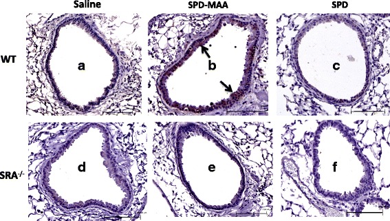
Fig. 6.

Representative lung tissue sections immunohistochemically stained for SPD-MAA. Immunohistochemical staining of SPD-MAA in lung airways of both WT and SR-A1 KO mice treated intranasally with saline, SPD-MAA (50 μg/mL) and SPD (50 μg/mL) for 3 weeks. A representative 4–5 μm-thick section of one mouse per treatment group is shown (20 × magnification). WT Saline a WT SPD-MAA b WT SPD c and SR-A1 KO Saline d, SR-A1 KO SPD-MAA e, SR-A1 KO SPD f. Line scale represents approx. 100 μm. Arrow denotes the staining and binding of MAA adduct
