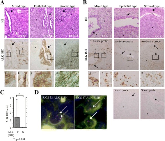
Fig. 1.

Full-length ALK expression in UCSs. a Staining by HE and IHC for ALK. Note the strong cytoplasmic ALK immunoreactivity in both carcinomatous (indicated by a and arrow) and sarcomatous components (indicated by b and asterisk) (indicated by closed boxes and magnified in the insets) in UCS51 (mixed type), UCS47 (epithelial type), and UCS33 cases (stromal type). Original magnification, ×100 and × 400 (inset). b Staining by HE and ISH for ALK mRNA. Note the mRNA signals in carcinomatous (a) and sarcomatous cells (b) (indicated by closed boxes and magnified in the insets) in UCS35 (mixed type), UCS29 (epithelial type), and UCS37 cases (stromal type). Original magnification, ×100 and × 400 (inset). c ALK IHC score in ALK mRNA-positive (P) and −negative (N) UCS cases. The data shown are means ± SDs. d FISH analysis of UCS33 and UCS47 cases. The interphase nuclei of both cases indicate absence of ALK rearrangement, in which the red and green signals remain fused (arrows)
