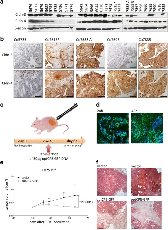
Fig. 4.

Analysis of claudin-3 and -4 expression and CPE sensitivity in patient derived colon carcinoma xenografts. a Western blot analysis for claudin-3 and claudin-4 expression, demonstrating highest expression of the claudins in colon cancer, since 26 of 27 analyzed tumors expressed claudin-3 and/or -4. Only one tumor (Co5735) did not express one of the claudins. b Representative images of immunohistochemistry for claudin-3 and -4 expression (brown staining) in the PDX tumors Co5735, Co7515*, Co7553A, Co7596 and Co7835, which correlated with protein expression (see A). The claudin-negative PDX Co5735 did not show any specific staining, whereas membranous localization of claudin-3 and/or -4 was found within the other representative PDX models. c Representative scheme of non-viral in vivo gene transfer using the jet-injection. d Intratumoral distribution of optCPE-GFP fusion protein expression 24 and 48 h after in vivo gene transfer of Co7515* tumors. Representative immunofluorescence images revealed strong optCPE-GFP accumulation within the tissues (green). Tissue disruption and cell death can be already seen after 24 h but strong areas of necrosis where detected after 48 h (see insets), indicating a successful gene transfer. Cell nuclei were counterstained with Dapi (blue). Scale bar: 100 μm. e Inhibition of Co7515* PDX tumor growth after optCPE-GFP gene transfer. The vector-transfected tumors serve as control. The in vivo optCPE-GFP gene transfer led to significant reduction in tumor growth (P = 0.0451); level of significance was determined by using non-parametric t-test; bars, S.E.M. f Representative H&E staining of terminal tumor sections (day 63 after Co7515* PDX inoculation) show incidence of massive necrotic areas in optCPE-GFP transfected tumors compared to vector control, indicating applicability of this gene therapy for colon carcinoma. Scale bar: 100 μm
