
Fig. 1.
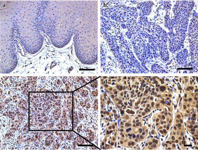
Immunohistochemical staining of SUZ12 in TSCC specimens and normal tongue mucosa. a Representative weak staining of SUZ12 (low expression) in normal human tongue mucosa (×200). Nuclei are counterstained with haematoxylin. b Representative weak staining of SUZ12 (low expression) in primary TSCC sample (×200). c Representative strong staining of SUZ12 (high expression) in primary TSCC sample (×200). d This image is magnified from the black box area in c (×400). SUZ12 expression is identified primarily in nuclei of cancer cells. Scale bar 100 μm