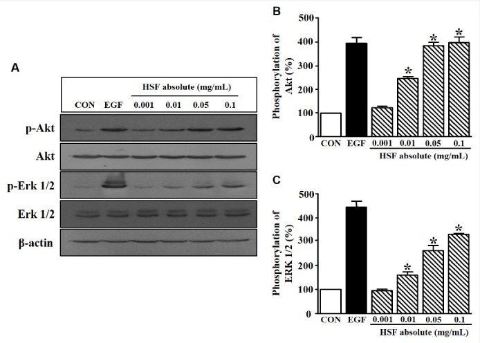
Figure 3.

Change in protein kinase level in Hibiscus syriacus L. flower (HSF) absolute-treated HaCaT cells. (a) HaCaT cells were seeded and incubated in serum-free medium for 24 h. The cells were then treated with recombinant human epidermal growth factor (EGF; 5 ng/mL) or HSF absolute (0.001–0.1 mg/mL) for 10 min. The cell lysates were immunoblotted with each kinase antibody (p-Akt, Akt, p-Erk 1/2, and Erk 1/2). (b and c), Statistical graphs were obtained from panel A. The basal level of p-Akt and p-Erk 1/2 in HaCaT cells in the quiescent state is expressed as 100% (n = 4). EGF was used as a positive control. Data are expressed as the mean ± SE. *P value less than 0.05 versus the nonstimulated group.
