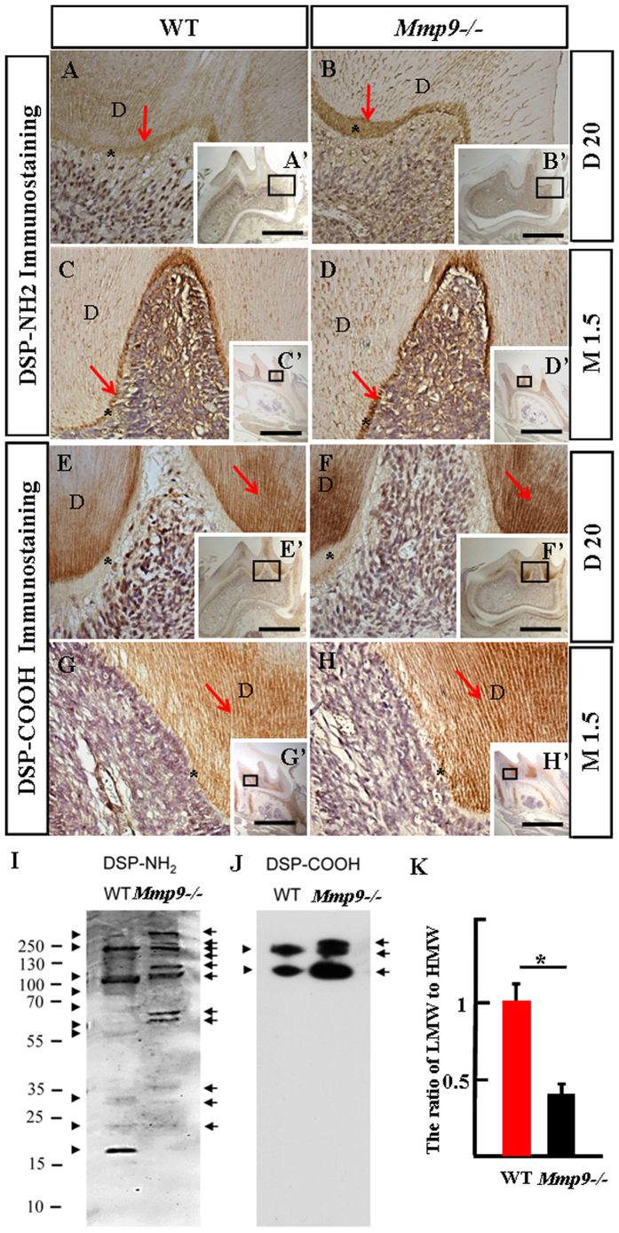
Figure 7. Immunolocalization and protein profiles of DSP in the Mmp9−/− and the wild-type molars.
(A–H, A’–H’) Immunodistribution of anti-DSP-NH2 antibody was strong in the predentin and the odontoblasts, but weak in the mineralized dentin. In contrast, DSP-COOH fragment(s) was strong in the mineralized dentin, but weak in the predentin and the odontoblasts at D20 and M1.5. (A–H) are higher magnification of the boxes in A’–H’. (I,J) Proteins were isolated from the wild-type and the Mmp9−/− teeth at D15, and Western blots with anti-DSP-NH2 and –COOH antibodies were performed. For normalization, the protein concentration should be measured and same amount of protein was loaded. Different DSP protein profiles were seen in the wild-type and the Mmp9−/− teeth. Arrows and arrowheads show DSP bands from the Mmp9−/− and the wild-type teeth, respectively. (K) The densitometry of anti-DSP-NH2 and -COOH bands of three independent experiments was performed and relative quantification was processed with the ImageJ software. The ratio of the LMW fragments (lower than 95 kDa) to the HMW DSP (higher than 95 kDa) was calculated. Statistical analysis was performed using Student’s t-test. *P < 0.05. In (A–I), *predentin; D, dentin. Scale bars: (A–H) 50 μm, (A’–H’) 1 mm.

