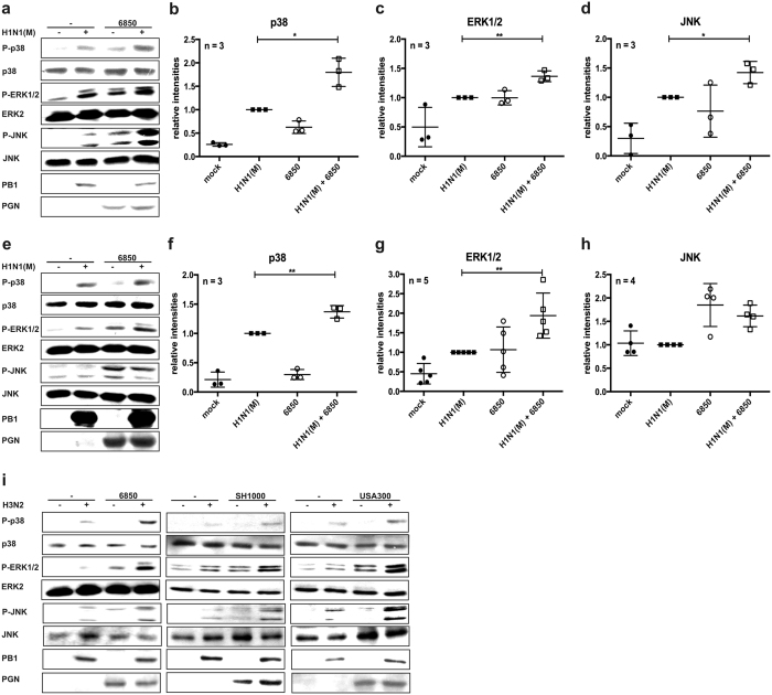
Figure 5. Phosphorylation of the MAPKs p38, ERK1/2 and JNK is increased upon IV/S. aureus super-infection.
Calu-3 (a–d) or A549 (e–i) cells were infected with IV H1N1(M) (a–h) or H3N2 (i) (MOI 5) for 0.5 h and super-infected with S. aureus 6850 (a–i), SH1000 or MRSA USA300 (i) (MOI 50). Extracellular bacteria were removed by gentamicin treatment 3 h after bacterial infection. At 8 h p.i. cells were lysed and whole cell lysates were subjected to Western blot analysis monitoring P-p38, P-ERK1/2, P-JNK, PGN and PB1. Equal protein loading was verified by detection of p38, ERK2 and JNK (a,e,i). Original blots are shown in Supplementary Fig. S8b (upper panel: Fig. 5a, lower panel: Fig. 5e) and S8c (upper panel: Fig. 5i [S. aureus 6850], lower panel [S. aureus SH1000, USA300]). Relative levels of P-p38, P-ERK1/2 and P-JNK compared to p38, ERK2 or JNK bands, respectively, were quantified by using ImageJ 2006.02.01 software (b–d,f–h). Means ± SD of at least three independent experiments are shown. Two-tailed unpaired t-tests were performed for comparison of IV H1N1(M)-infected and IV H1N1(M)/S. aureus 6850 super-infected samples (*p < 0.05, **p < 0.01).

