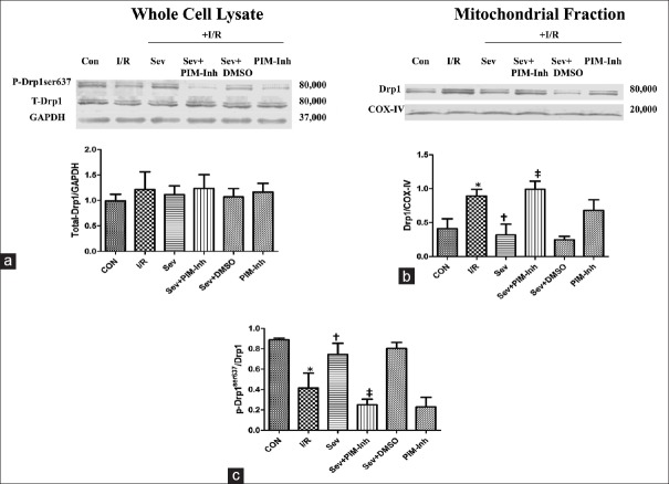
Figure 3.
Western blotting analysis of total Drp1 and p-Drp1ser637 in myocardium as well as Drp1 translation to the mitochondria. The total Drp1 (80,000 Da) (a) and the Drp1 in mitochondria (c) were analyzed by Western blotting with specific Drp1 antibody at 1 h of reperfusion; The p-Drp1ser637 (80,000 Da) (b) was analyzed by Western blotting with specific p-Drp1ser637 antibody at 1 h of reperfusion (n = 3 for each group). Data were reported as mean ± SD. *P < 0.05 versus CON; †P < 0.05 versus I/R; ‡P < 0.05 versus Sev (one-way ANOVA). CON: Control group; I/R: Ischemia/reperfusion; Sev: Sevoflurane postconditioning; Sev+PIM-Inh: Sevoflurane postconditioning + Pim-1 inhibitor II; Sev+DMSO: Sevoflurane postconditioning + dimethyl sulfoxide; PIM-Inh: Pim-1 inhibitor II; Drp1: Dynamics-related protein 1; GAPDH: Glyceraldehyde-3-phosphate dehydrogenase; COX-IV: Cytochrome C oxidase IV; SD: Standard deviation; ANOVA: Analysis of variance.

