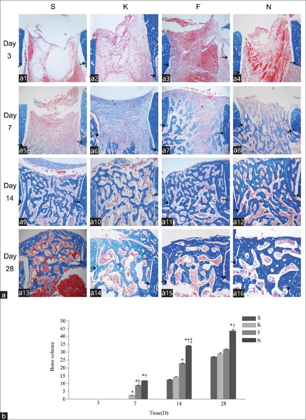
Figure 2.
Effect of 15d-PGJ2 on new bone formation in the cortical defect area. (a) The new bone formation was examined in specimens from animals treated with saline (a1, a5, a11 and a13), empty nanocapsules (a2, a6, a10 and a14), free 15d-PGJ2 (a3, a7, a11 and a15), and 15d-PGJ2-NC (a4, a8, a12 and a16) on days 3 (a1-a4), 7 (a5-a8), 14 (a9-a12), and 28 (a13-a16) after surgery, respectively. Black arrows indicated the margin of the surgically created cortical bone wound (Masson's Trichrome, original magnification ×100). (b) Semi-quantitative evaluation of bone formation in the cortical defect area. Three sections from each group were obtained on days 3, 7, 14, and 28. Values represent the mean ± standard deviation of three sections. *P < 0.05 versus groups S; †P < 0.05 versus groups K; ‡P < 0.05 versus groups F. 15d-PGJ2-NC: 15-Deoxy-∆12,14-prostaglandin J2 nanocapsules; S: Saline; K: Empty nanocapsules; F: Free 15-Deoxy-∆12,14-prostaglandin J2; N: 15-Deoxy-∆12,14-prostaglandin J2 nanocapsules.

