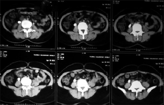
Figure 6.

Computed tomography kidneys, ureters, bladder (P + C) of patient with right hydroureter and right ureterovaginal fistula

Computed tomography kidneys, ureters, bladder (P + C) of patient with right hydroureter and right ureterovaginal fistula