
Fig. 5.
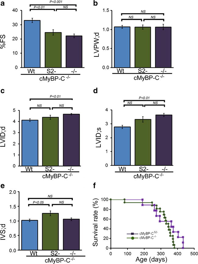
M-mode echocardiography indices of LV end-diastolic diameter and function. a Percent fractional shortening (%FS). b LV diastolic posterior wall thickness. c LV diastolic volume (LV vol;d). d LV systolic volume (LV vol;s). e Diastolic interventricular septum thickness (IVS;d). (n ≧ 10 mice per group, mean ± S.E.M). P value versus cMyBP-CWt mice by Tukey’s post hoc test. All measurements were carried out with 3-month-old mice. f Kaplan-Meier analysis of survival probabilities for the null (n = 16) versus the cMyBP-CS2− (n = 9) animals. There were no statistically significant differences between the two curves